

This is part three of a five-part series decoding the biological evidence behind five of the most dismissed symptoms in Long COVID, ME/CFS, PoTS and related conditions. Part one looked at brain fog and the PET scan evidence of dopamine system injury. Part two looked at the PEM crash and the muscle biopsy findings that make post-exertional malaise impossible to dismiss. Today: the heart rate spike.
Of all the symptoms in this space, postural orthostatic tachycardia syndrome attracts a particular kind of dismissal. The racing heart, the lightheadedness, the inability to stand without symptoms. These are routinely attributed to anxiety, deconditioning, or habit. Patients are told their nervous system is overreacting. They are told to exercise more. They are told to drink more water and push through.
The studies below describe a different picture. A picture of autoantibodies targeting autonomic receptors, of measurable reductions in blood flow to the brain on standing, of structural damage to the small nerve fibres that regulate blood vessels and of a vascular system that is not pooling blood because of weakness or fear but because of specific, identifiable physiological failure. These are not hypotheses. They are findings, in peer-reviewed journals, from researchers who measured what is actually happening in PoTS bodies.
Study 1: The autoantibodies attacking autonomic receptors
Gomez-Esteban JC, et al. Involvement of autoantibodies against G protein-coupled receptors in post-COVID condition and Chronic Fatigue Syndrome. Scientific Reports. 2026;16. https://doi.org/10.1038/s41598-026-49131-9
Published in Scientific Reports in May 2026, this study examined autoantibodies targeting adrenergic and muscarinic receptors, the receptors that regulate heart rate, blood vessel tone and the autonomic nervous system, in 96 post-COVID patients, 59 ME/CFS patients and 36 healthy controls.
The adrenergic system is the mechanism by which the body controls the cardiovascular response to posture change. Beta-2 adrenergic receptors on blood vessel walls respond to norepinephrine to regulate vascular tone. When autoantibodies target these receptors, they disrupt the normal regulatory signal, interfering with the precisely timed vascular response that keeps blood pressure and heart rate stable when you move from lying to standing.
ME/CFS patients in this study showed significantly elevated beta-2 adrenergic autoantibody titers compared to both post-COVID patients and healthy controls (p = 0.046). In post-COVID patients, M3 muscarinic receptor antibodies were elevated. Critically, in the post-COVID group, beta-2 antibody levels correlated directly with autonomic dysfunction scores (r = 0.27, p = 0.048). Higher antibody levels meant worse autonomic dysregulation.
This is not an anxiety response. Anxiety does not produce elevated beta-2 adrenergic autoantibody titers. The immune system attacking its own autonomic receptors does.
Study 2: The brain losing blood when you stand up
van Campen CLMC, Visser FC, Rowe PC. Cerebral blood flow is reduced in ME/CFS during head-up tilt testing even in the absence of hypotension or tachycardia. Clinical Neurophysiology Practice. 2020;5:50-58. https://doi.org/10.1016/j.cnp.2020.04.003
This study, from researchers at Stichting CardioZorg in the Netherlands, used Doppler imaging of the carotid and vertebral arteries to measure total cerebral blood flow in 429 ME/CFS patients and 44 healthy controls during a 30-minute head-up tilt test.
The finding is stark. Healthy controls showed a 7% reduction in cerebral blood flow on tilting, the normal physiological response. ME/CFS patients showed a 26% reduction overall. That figure held across subgroups: 24% in those with a normal heart rate and blood pressure response, 28% in those with delayed orthostatic hypotension and 29% in those with a PoTS-pattern tachycardia. Ninety percent of ME/CFS patients exceeded the abnormal threshold of a 13% reduction, two standard deviations below the healthy control mean.
What this means in practice is that when many PoTS and ME/CFS patients stand up, their brain is receiving roughly a quarter less blood than it should. The lightheadedness, the cognitive blunting, the visual changes, the overwhelming need to sit or lie down. These are not sensations without a substrate. The substrate is a brain running significantly below its required blood supply.
The study also reveals why standard tilt table tests miss this. Most clinical tilt tests measure heart rate and blood pressure. They do not measure cerebral blood flow. A patient can have a 26% drop in cerebral perfusion with a normal heart rate and blood pressure reading. Normal on paper. Not normal in the brain.
Study 3: The nerve fibres that regulate blood vessels are damaged
Novak P, et al. Small fiber neuropathy in Long COVID, ME/CFS, and hypermobile Ehlers-Danlos syndrome. PLOS ONE. 2026. https://doi.org/10.1371/journal.pone.0341278
Small fiber neuropathy is damage to the thinnest nerve fibres in the peripheral nervous system. These fibres have two relevant functions: transmitting sensory signals alongside regulating the autonomic function of blood vessels and sweat glands. When they are damaged, the body loses part of its capacity to automatically adjust vascular tone in response to posture, which is precisely the mechanism that fails in PoTS.
This 2026 study from Peter Novak at Brigham and Women’s Hospital, Harvard, examined 143 Long COVID patients, 170 ME/CFS patients, 290 patients with hypermobile Ehlers-Danlos syndrome and 73 healthy controls, using both structural markers (epidermal nerve fibre density from skin punch biopsies) and functional markers including sudomotor testing.
Approximately 80 to 90 percent of patients across these conditions showed evidence of small fiber neuropathy using combined criteria. Abnormal epidermal nerve fibre density was found in 48% of Long COVID patients and 34% of ME/CFS patients. Sudomotor dysfunction, reflecting damage to the autonomic nerve fibres supplying sweat glands, which share the same small fiber pathway as the vascular autonomic nerves, was present in both groups at rates significantly exceeding healthy controls.
In a subset of PoTS patients, the damage is specifically to the small fibres innervating blood vessel walls in the lower limbs. When these fibres cannot transmit the vasoconstriction signal on standing, blood pools. The heart rate rises to compensate. This is PoTS with a structural cause visible on biopsy.
Study 4: The blood pooling measured in millilitres, and the blood volume that is genuinely reduced
Kulapatana P, et al. Blood volume deficit in postural orthostatic tachycardia syndrome assessed by semiautomated carbon monoxide rebreathing. Clinical Autonomic Research. 2025. https://doi.org/10.1007/s10286-024-01091-8
The excessive blood pooling in PoTS and the low circulating blood volume that compounds it, are among the most replicated findings in dysautonomia research. Tachycardia on standing is the symptom. The blood distribution failure underneath it is the cause.
This 2025 study measured blood volume directly using carbon monoxide rebreathing in 21 female PoTS patients and 19 healthy female controls. PoTS patients had blood volumes approximately 14% below normal. Total blood volume was 64.53 mL/kg in patients versus 76.78 mL/kg in controls. Plasma volume was 39.50 mL/kg versus 48.20 mL/kg. Red blood cell volume was reduced by a comparable margin. There was a strong negative correlation between blood volume deficit and upright heart rate, meaning the lower the blood volume, the higher the compensatory tachycardia.
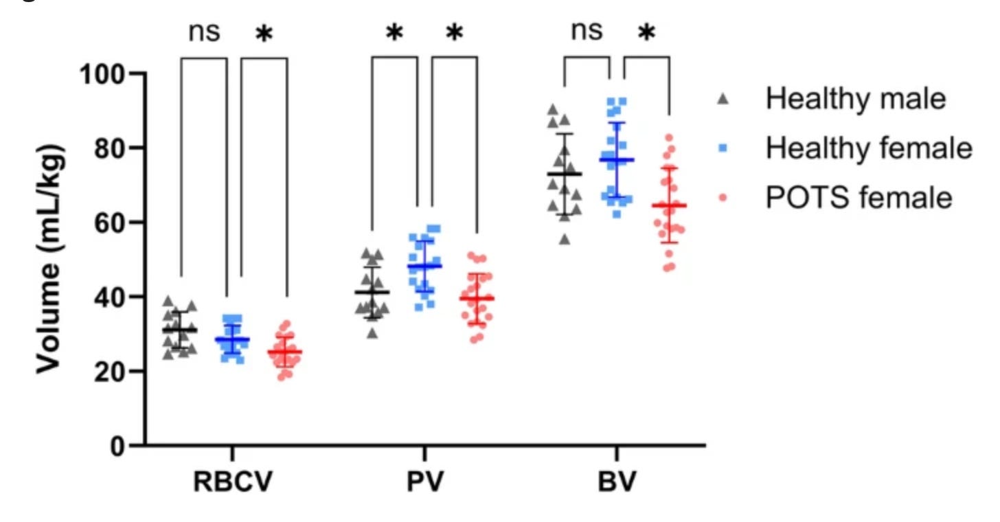
The tachycardia that defines PoTS diagnostically is the cardiovascular system’s response to a real, physical problem: not enough blood returning to the heart because there is genuinely less of it in circulation, pooling in the wrong places. The body is not overreacting. It is compensating for an actual shortfall that is measurable in millilitres.
Study 5: The immune and inflammatory signature that separates PoTS from anxiety
Seeley MA, Stavrakis S, et al. Comparative Analysis of Circulating Cytokines and Adrenergic Autoantibodies in Postural Orthostatic Tachycardia Syndrome, Postacute Sequelae of SARS-CoV-2, and Healthy Controls. Journal of the American Heart Association. 2026;15(11). https://pubmed.ncbi.nlm.nih.gov/42179241/
This 2026 study from the Journal of the American Heart Association compared cytokine profiles in 24 PoTS patients, 24 post-acute sequelae of SARS-CoV-2 patients and 19 healthy controls. PoTS patients showed significantly lower interleukin-2 and elevated interleukin-8 compared to controls, along with elevated tumour necrosis factor-alpha.
Interleukin-2 is a cytokine associated with T cell regulation and immune homeostasis. Its reduction in PoTS is consistent with the broader pattern of immune dysregulation seen across post-viral autonomic conditions. Elevated IL-8 is a pro-inflammatory signal linked to neutrophil recruitment. Elevated TNF-alpha is a hallmark of systemic inflammatory activation.
These are not the cytokine profiles of people who are anxious about standing up. Anxiety does not selectively reduce interleukin-2 or elevate tumour necrosis factor-alpha. A dysregulated immune system responding to a post-viral process does.
The study also noted that cytokine and autoantibody measures alone did not predict PoTS in every patient, a finding consistent with the understanding that PoTS is a heterogeneous condition with multiple converging mechanisms, of which autoimmunity and inflammation are significant pathways rather than a single universal cause. Not every patient has identical biology. But every patient has a biology. And it is measurable.
What this means
Five findings. Autoantibodies targeting the adrenergic receptors that regulate cardiovascular response to posture. A 26% reduction in cerebral blood flow on standing, visible on Doppler imaging, in 90% of patients. Structural damage to the small nerve fibres that maintain vascular tone, found in biopsies in 80 to 90% of patients with related conditions. Quantifiable blood pooling and a plasma volume roughly 14% below normal in PoTS patients, confirmed by direct measurement. An inflammatory cytokine signature that distinguishes PoTS patients from healthy controls at the immune level.
None of these findings are consistent with the explanation that PoTS is anxiety, deconditioning, or a learned avoidance of standing. All of them are consistent with an autonomic nervous system that has been structurally and immunologically disrupted, producing a measurable, reproducible failure of the cardiovascular system to handle orthostatic stress normally.
The question, as always, is how long it takes that evidence to reach the clinical settings where patients are still being told to push through.
The views and opinions expressed in Dysautonomia Decoded are my own and do not represent those of my employer or any affiliated organisation.
References
Gomez-Esteban JC, et al. Involvement of autoantibodies against G protein-coupled receptors in post-COVID condition and Chronic Fatigue Syndrome. Scientific Reports. 2026;16. https://doi.org/10.1038/s41598-026-49131-9
van Campen CLMC, Visser FC, Rowe PC. Cerebral blood flow is reduced in ME/CFS during head-up tilt testing even in the absence of hypotension or tachycardia. Clinical Neurophysiology Practice. 2020;5:50-58. https://doi.org/10.1016/j.cnp.2020.04.003
Novak P, et al. Small fiber neuropathy in Long COVID, ME/CFS, and hypermobile Ehlers-Danlos syndrome. PLOS ONE. 2026. https://doi.org/10.1371/journal.pone.0341278
Kulapatana P, Diedrich A, et al. Blood volume deficit in postural orthostatic tachycardia syndrome assessed by semiautomated carbon monoxide rebreathing. Clinical Autonomic Research. 2025. https://doi.org/10.1007/s10286-024-01091-8
Seeley MA, Stavrakis S, et al. Comparative Analysis of Circulating Cytokines and Adrenergic Autoantibodies in Postural Orthostatic Tachycardia Syndrome, Postacute Sequelae of SARS-CoV-2, and Healthy Controls. Journal of the American Heart Association. 2026;15(11). https://pubmed.ncbi.nlm.nih.gov/42179241/
I wish I could love this a million times and send it to everyone I know (some of them, undoubtedly, would still accuse me of being dramatic, but I swear...it's always been real..."you can't fake a tilt table test"...I whisper to myself for the millionth time).
Loved this as always, thank you so much for your work