

Today let's decode a brain imaging study published in eBioMedicine in July 2026 by researchers at the Centre for Addiction and Mental Health in Toronto that has been circulating widely in the Long COVID research community this week. It provides what the authors describe as the strongest evidence to date that Long COVID is associated with injury to dopamine-releasing neurons in the brain. The finding matters for several reasons: it provides a specific, measurable, imaging-based biological explanation for some of Long COVID's most disabling symptoms, it connects to a known mechanism from other neurological conditions, and it is already informing the design of a clinical trial.
What the dopamine system does
Dopamine is one of the brain’s primary neurotransmitters, a chemical messenger that carries signals between neurons. It is involved in motivation, movement, memory consolidation, reward processing, along with the regulation of attention. When people talk about dopamine they often think of pleasure, but its functions are considerably broader and more fundamental than that.
The striatum is the brain structure most densely populated with dopamine-releasing neurons. It sits deep in the brain and plays a central role in initiating and regulating movement, sustaining motivation, along with supporting working memory and executive function. Damage to dopamine-releasing neurons in the striatum is what causes the movement slowness, rigidity and cognitive symptoms of Parkinson’s disease. It is also associated with the profound fatigue and motivational impairment seen in depression and other neuropsychiatric conditions.
The CAMH researchers asked a specific question: are Long COVID patients showing signs of the same kind of dopamine neuron injury, and if so, does it correlate with the severity of their symptoms?
What the study did
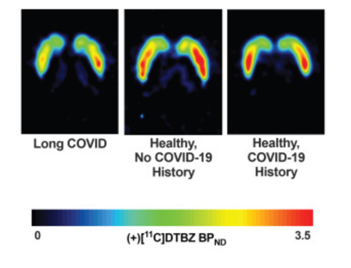
The study enrolled 24 adults with Long COVID, defined as symptoms persisting for more than three months following confirmed COVID-19 infection, alongside 24 age-matched healthy controls. The comparison group was extended to 43 participants for exploratory analyses. All participants underwent PET brain imaging using a radiotracer called VMAT2, which binds to vesicular monoamine transporter 2, a protein found on the surface of dopamine nerve terminals. VMAT2 binding is a well-established marker of dopamine neuron integrity. Lower VMAT2 binding indicates reduced dopamine nerve terminal density, which reflects injury to or loss of dopamine-releasing neurons.
The study ran from 2022 to 2025 in Toronto, making it a properly prospective study rather than a retrospective analysis of existing data.
What they found
Long COVID patients showed significantly lower VMAT2 binding than healthy controls across all major regions of the striatum. This was not a marginal or borderline finding. The reduction was consistent across the ventral striatum, the dorsal putamen, and the caudate putamen, three distinct striatal subregions with different functional roles.
The correlations between regional VMAT2 reductions and specific symptom domains were equally striking. Reduced binding in the ventral striatum correlated with loss of motivation and fatigue. Reduced binding in the dorsal putamen correlated with slowed movement. Reduced binding in the caudate putamen correlated with memory difficulties. Each subregion maps onto a different symptom, and each correlation was statistically significant.
This specificity is important. It is not simply that people with Long COVID have generally disrupted brain function. The damage appears to be regionally specific, with different parts of the dopamine system showing reductions that correspond to different parts of the symptom profile.
The findings build on earlier work by the same research team demonstrating that people with Long COVID have unusually high levels of brain inflammation on PET imaging. The new study suggests a mechanism connecting those earlier findings: inflammation in the brain may be causing direct injury to dopamine-releasing neurons, which then produces the motivational, cognitive and motor symptoms characteristic of Long COVID neurological presentation.
Why this matters for understanding Long COVID
The symptom cluster of fatigue, brain fog, lack of motivation, slowed thinking and memory impairment is among the most consistently reported and most consistently dismissed features of Long COVID. Patients are frequently told these symptoms are secondary effects of deconditioning, anxiety, or simply not being well. The CAMH study provides imaging evidence that they reflect specific, measurable biological injury to a specific brain system.
The relevance to ME/CFS is equally significant. Reduced dopamine neurotransmission has been proposed as a mechanism in ME/CFS for years, largely on the basis of indirect evidence from symptom profiles along with some cerebrospinal fluid studies. A PET imaging study of the kind conducted here has not previously been done in ME/CFS, but the symptom overlap is compelling enough that the findings are likely to prompt similar investigations.
The connection to neuroinflammation is also worth emphasising. The same CAMH group previously found elevated neuroinflammation markers in Long COVID using PET imaging. The dopamine findings now suggest a downstream consequence of that inflammation: when the brain is chronically inflamed, dopamine neurons are damaged. The inflammatory origin of the dopamine injury matters therapeutically because it means that treatments targeting neuroinflammation might also protect or restore dopamine function.
What happens next
Dr Jeffrey Meyer, senior author of the study and senior scientist at CAMH’s Brain Health Imaging Centre, is moving directly from this observational finding to a clinical trial. The planned trial, in collaboration with University Health Network, will test dopamine-targeting treatments for fatigue and memory problems in Long COVID patients. The research group has already applied for a patent for rasagiline, a drug used in Parkinson’s disease that inhibits the breakdown of dopamine, as a potential Long COVID treatment. A tyramine-based approach to increasing dopamine precursors is also being explored.
It is worth being clear about what rasagiline is and is not. It is an approved medication used in Parkinson’s disease, where it slows the progression of dopamine neuron loss. Whether the same drug, used in a different disease at a potentially different stage of injury, will produce meaningful clinical benefit in Long COVID patients is not established. The trial is necessary to answer that question. Until results are available, this is a hypothesis supported by compelling imaging evidence, not a treatment recommendation.
The limitations worth naming
Twenty-four Long COVID patients is a small sample. The findings need to be replicated in a larger cohort before they can be considered definitive. The study was conducted at a single centre in Toronto, and the Long COVID population attending a Canadian research centre may not be representative of all Long COVID presentations globally. The cross-sectional design means the study captures a snapshot rather than tracking how dopamine neuron density changes over time in the same individuals. It cannot tell us whether the damage is progressive, stable, or reversible.
The correlation between VMAT2 reductions and specific symptom domains, while statistically significant, does not establish that reduced dopamine function causes those symptoms. It is possible that both the dopamine injury and the symptoms are downstream of a third factor, most plausibly the neuroinflammation documented in the earlier CAMH work.
The take home message
Researchers scanned the brains of Long COVID patients and found significantly reduced dopamine nerve terminal density across all major regions of the striatum. The reductions correlated specifically with fatigue, motivational impairment, slowed movement and memory difficulty. The damage is consistent with the kind of injury seen in other conditions where dopamine neuron loss produces neurological symptoms. A clinical trial of dopamine-targeting treatment is being planned.
Brain fog, fatigue and lack of motivation in Long COVID are not unexplained. They are visible on a brain scan. They correlate with specific patterns of injury in a specific brain system. The biology is there. The question is now what to do about it.
The views and opinions expressed in Dysautonomia Decoded are my own and do not represent those of my employer or any affiliated organisation.
References
Meyer JH, et al. Loss of vesicular monoamine transporter 2 in striatum of long COVID and relationship to neuropsychiatric symptoms. eBioMedicine. 2026;130:106339. https://doi.org/10.1016/j.ebiom.2026.106339
Centre for Addiction and Mental Health. New study provides first evidence of dopamine system injury in the brain of long COVID patients. EurekAlert. July 10 2026. https://www.eurekalert.org/news-releases/1134884
Thanks for sharing this, fascinating as always
Thanks for sharing this really interesting topic. As a pharmacist, I find the discussion around dopamine regulation, nutrient cofactors, and methylation especially relevant—so many people overlook how these metabolic pathways can influence mood and motivation. Looking forward to more of your insights.