


Today let’s decode a study that took an unusual route to understanding what Long COVID and ME/CFS do to the body’s smallest blood vessels. Rather than invasive biopsies or expensive imaging, researchers at the Technical University of Munich looked into patients’ eyes. Specifically at the tiny blood vessels in the retina, which share their embryological origin, structure and regulatory mechanisms with the blood vessels of the brain. What you see in the retina, it turns out, can reflect what is happening in the cerebral microcirculation.
Why the retina
The retina is the only place in the body where blood vessels can be observed non-invasively, in real time, without contrast agents or radiation. Retinal vessel analysis measures the diameter, function and response of these tiny vessels, and decades of cardiovascular research have established that retinal microvascular changes reliably predict systemic microvascular disease. Changes in retinal vessel diameter and function predict stroke risk, cardiovascular mortality and kidney disease with meaningful accuracy. The retina is essentially a window into the vascular health of the whole body, particularly the brain.
This is directly relevant to Long COVID and ME/CFS because persistent symptoms in both conditions, fatigue, brain fog, cognitive impairment, autonomic dysfunction, are increasingly linked to what is happening in the brain’s small blood vessels. If the brain is not getting enough blood, or if the blood vessels regulating its supply are not functioning properly, the symptoms that result look very much like the ones these patients experience.
What the All Eyes on PCS study actually did
The study, published in BMC Medicine in August 2026 by Wallraven, Günthner and colleagues at the Technical University of Munich and Charité Berlin, recruited 102 post-COVID syndrome patients and compared them with 204 age and sex matched healthy controls. This is a prospective, observational case-control design, which is a substantially stronger evidence base than the retrospective or self-report studies that dominate this space.
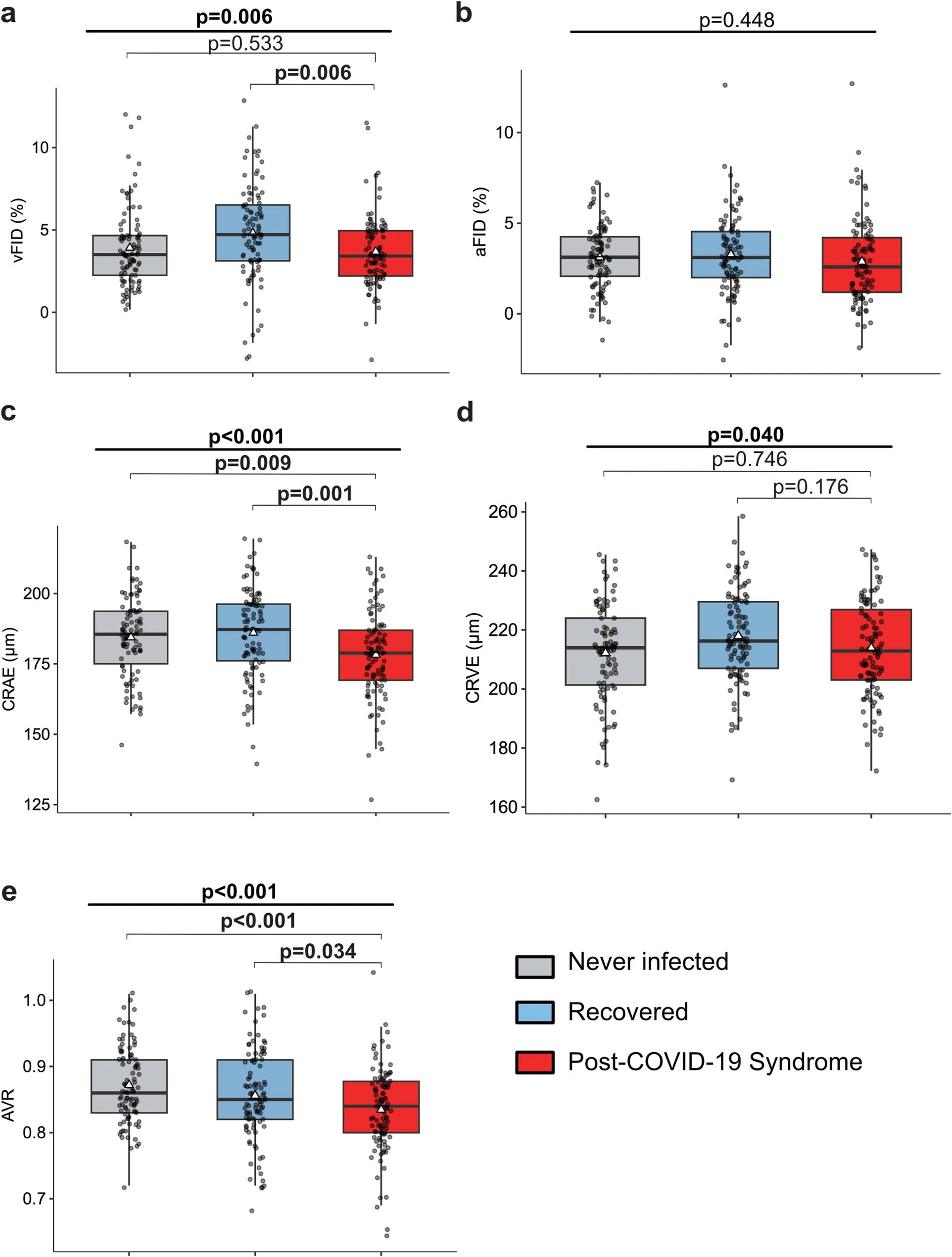
Participants underwent retinal vessel analysis measuring both static parameters (the diameter of arterioles and venules) and dynamic parameters (how vessels dilate in response to flickering light, a standardised stimulus called flicker light-induced retinal vasodilation). They also had blood tests for inflammatory and endothelial dysfunction markers including von Willebrand factor, soluble thrombomodulin and various cytokines. A subset of 20 patients with ME/CFS were also included for comparison.
What they found
Post-COVID patients showed significantly impaired retinal vessel dilation in response to flickering light compared to healthy controls. This is the dynamic functional measure, not just the structural diameter, and it reflects the ability of blood vessel walls to dilate appropriately in response to increased demand. Impaired retinal vessel dilation in this test is a marker of endothelial dysfunction, meaning the cells lining the inside of the blood vessels are not responding properly.
The severity of retinal microvascular changes correlated significantly with symptom severity. Patients with more severe post-COVID symptoms showed worse retinal vessel function than those with milder presentations. Neurocognitive symptoms including fatigue, impaired concentration and brain fog were specifically linked to worse retinal microvascular markers, consistent with the hypothesis that endothelial dysfunction contributes directly to the central nervous system manifestations of post-COVID syndrome.
Circulating markers of endothelial dysfunction and inflammation were elevated in post-COVID patients compared to healthy controls, and these markers correlated with both symptom severity and the retinal findings.
The ME/CFS subgroup showed similar retinal microvascular changes to post-COVID patients, suggesting shared underlying mechanisms. This is consistent with the Harvard comparison study published earlier in 2026, which found that Long COVID and ME/CFS patients were essentially indistinguishable on detailed autonomic testing.
The microclotting connection
These findings sit within a larger picture of microvascular dysfunction in post-COVID and ME/CFS that has been building in the research literature over the past four years. Professor Etheresia Pretorius at Stellenbosch University and colleagues have documented fibrin amyloid microclots in the blood of Long COVID patients, tiny, abnormally resistant clots that may impede blood flow through the smallest vessels. A 2024 analysis of ME/CFS plasma found dysregulated coagulation, endothelial dysfunction and downregulation of complement machinery. The Wallraven study adds retinal microvascular evidence to this picture, showing that the endothelial dysfunction is not just detectable in blood markers but visible in the functioning of real blood vessels in real patients.
Put together, the emerging picture is of a post-viral state in which the cells lining blood vessel walls throughout the body, and particularly in the brain’s microvasculature, are failing to function normally. Blood vessel dilation is impaired. Inflammatory markers are elevated. Coagulation is dysregulated. And these changes are detectable years after the initial infection in patients who had a relatively mild acute illness.
What this might mean for treatment
The authors note that endothelial function is a potential therapeutic target. Several interventions that improve endothelial function in other contexts, including exercise in those who can tolerate it, certain medications used in cardiovascular disease, and anti-inflammatory approaches, are already being studied in Long COVID and ME/CFS. The retinal vessel analysis framework could provide a non-invasive, accessible way of tracking whether those interventions are actually improving microvascular health, rather than relying solely on subjective symptom reports.
A separate 2024 study found that 41% of post-acute COVID syndrome patients had microvascular dysfunction detectable over two years after a mild initial infection, and that its presence was associated with increasing NT-proBNP levels over time, a marker of cardiovascular stress and potential cardiac remodeling. The scale and persistence of the vascular problem is becoming increasingly hard to dismiss.
The limitations worth naming
This is a single-centre study conducted in Munich, and the patient population attending a specialist post-COVID clinic may not be representative of all Long COVID presentations. The ME/CFS subgroup was small at 20 patients, limiting the conclusions that can be drawn from that comparison. The cross-sectional nature of the main analysis means it captures a snapshot rather than tracking how vascular function changes over time. And retinal vessel analysis, while validated, requires specialist equipment and trained operators that are not widely available in routine clinical settings.
The study also cannot yet tell us whether the endothelial dysfunction causes symptoms, is a consequence of other processes, or is one component of a multi-system dysfunction in which cause and effect are deeply entangled.
The take home message
Researchers looked into the eyes of Long COVID patients and found that the tiny blood vessels in their retinas are not functioning properly, in ways that correlate directly with the severity of their symptoms. The same findings appear in ME/CFS. The changes are detectable years after a mild infection. And they point toward the blood vessel lining itself as a potential site of disease and a potential target for treatment.
This is not in the mind. It is in the microvasculature.
The views and opinions expressed in Dysautonomia Decoded are my own and do not represent those of my employer or any affiliated organisation.
References
Wallraven T, Günthner R, Lethen I, et al. Microvascular remodeling and endothelial dysfunction across the post-COVID-19 spectrum: a prospective observational case-control study. BMC Medicine. 2026;24(1):456. https://doi.org/10.1186/s12916-026-05144-9
Nunes M, Vlok M, Proal A, Kell DB, Pretorius E. Data-independent LC-MS/MS analysis of ME/CFS plasma reveals a dysregulated coagulation system, endothelial dysfunction, downregulation of complement machinery. Cardiovascular Diabetology. 2024;23:254. https://doi.org/10.1186/s12933-024-02315-x
Novak P, Systrom DM, et al. Shared autonomic phenotype of Long COVID and ME/CFS. PLOS ONE. 2026;21(1):e0341278.
American Journal of Medicine. Post-acute COVID-19 syndrome: prevalence of peripheral microvascular endothelial dysfunction and associations with NT-proBNP dynamics. 2024. https://doi.org/10.1016/j.amjmed.2024.09.022
The observation of fibrin amyloid blood clots in patients with long covid is remarkable. The first time I am hearing about amyloid outside the context of Alzheimer's disease.
What other uses are there for amyloid, beyond what appears to be happening here is protecting the body from whatever long covid is doing?
Are there any connections between the pathophysiology of long covid and Alzheimer's disease, beyond the fact that both of them can modify the brain and reduce its function?
This study and further research based on this has the opportunity to shed light on not only long covid but other seemingly unrelated diseases like Alzheimer's and diabetic retinopathy which can affect retinal blood vessels.
It's all in the eye of the beholder.