


If you have a chronic illness such as PoTS, ME/CFS, Long COVID, EDS or MCAS, the chances are high that you have been told at some point that your symptoms are anxiety, or something you could think your way out of. The research below will not undo the harm of that experience. But it does demonstrate that each of these conditions leaves a measurable, objective, biological footprint that is visible to anyone willing to look.
These are not opinion pieces or patient surveys. They are peer-reviewed studies published in some of the most rigorous journals in the world. Each one found something that should not exist in a condition that is “all in your head.”
1. PoTS: The Brain Is Visibly Running Low on Blood
Seeley MC, et al. Novel brain SPECT imaging unravels abnormal cerebral perfusion in patients with postural orthostatic tachycardia syndrome and cognitive dysfunction. Scientific Reports. 2025;15:3487.
Researchers at the University of Adelaide used brain SPECT imaging, a nuclear medicine scan that measures regional cerebral blood flow, in 56 PoTS patients who reported cognitive symptoms. The results were unambiguous: 61% showed abnormal blood flow patterns in the brain, including while lying flat. The regions most affected were the prefrontal cortex and sensorimotor cortex, the areas responsible for attention, decision-making, and memory.
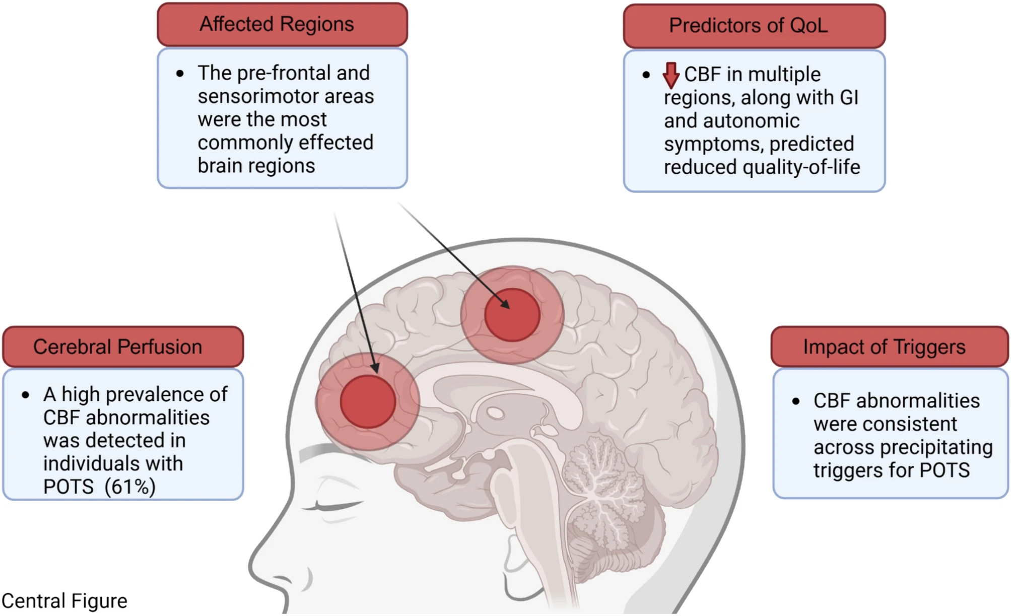
When the researchers modelled what was actually driving health and quality of life in these patients, the number of brain regions with abnormal blood flow accounted for 51% of the variance. Standing heart rate, the number used to diagnose PoTS, accounted for none of that variance independently.
Brain fog in PoTS is not a secondary symptom or a psychological response to feeling unwell. It is measurable, visible on imaging, directly linked to reduced cerebral perfusion, and present even before the patient stands up. The brain is not getting enough blood, and the consequences of that are showing up on a scan.
2. ME/CFS: A Protein Is Physically Dismantling the Mitochondria
Wang PY, Ma J, Kim YC, et al. WASF3 disrupts mitochondrial respiration and may mediate exercise intolerance in myalgic encephalomyelitis/chronic fatigue syndrome. Proceedings of the National Academy of Sciences. 2023;120(34):e2302738120.
Researchers at the National Heart, Lung and Blood Institute at the NIH began investigating the cells of a single ME/CFS patient whose muscles were consistently failing to produce energy at a normal rate. What they found was specific: a protein called WASF3 was present at abnormally high levels in her skeletal muscle, and it was physically disrupting the assembly of respiratory supercomplexes, the protein structures that mitochondria use to produce ATP, the body’s energy currency.
WASF3 is produced in excess when cells are under endoplasmic reticulum stress. In this patient, it was travelling to the mitochondria and preventing the machinery from assembling properly. The cell’s power plant was being actively sabotaged by one of its own proteins.
The researchers then examined skeletal muscle biopsies from a broader cohort of ME/CFS patients and found elevated WASF3 levels across the group, not just in the index case. Crucially, blocking ER stress pharmacologically reduced WASF3 levels and restored mitochondrial function in the patient’s cells. This is the most specific molecular mechanism identified in ME/CFS to date: a named protein, a named target, a named consequence, visible in biopsies from multiple patients, with a pharmacological reversal demonstrated in cell models.
3. Long COVID: The Crash Is Visible Under a Microscope
Appelman B, Charlton BT, Goulding RP, et al. Muscle abnormalities worsen after post-exertional malaise in long COVID. Nature Communications. 2024;15:17.
This is perhaps the single most viscerally compelling study in the post-viral illness literature of the past five years. Researchers took muscle biopsies from 25 Long COVID patients with post-exertional malaise before exercise and one day after exercise triggered a crash, running the identical protocol in 21 healthy controls.
Before a single exercise bout, Long COVID muscle was already different. Mitochondrial enzyme activity was reduced. The proportion of fast-twitch fibres that fatigue quickly was higher. These differences were present at baseline, without any exercise having occurred. The starting line was already behind
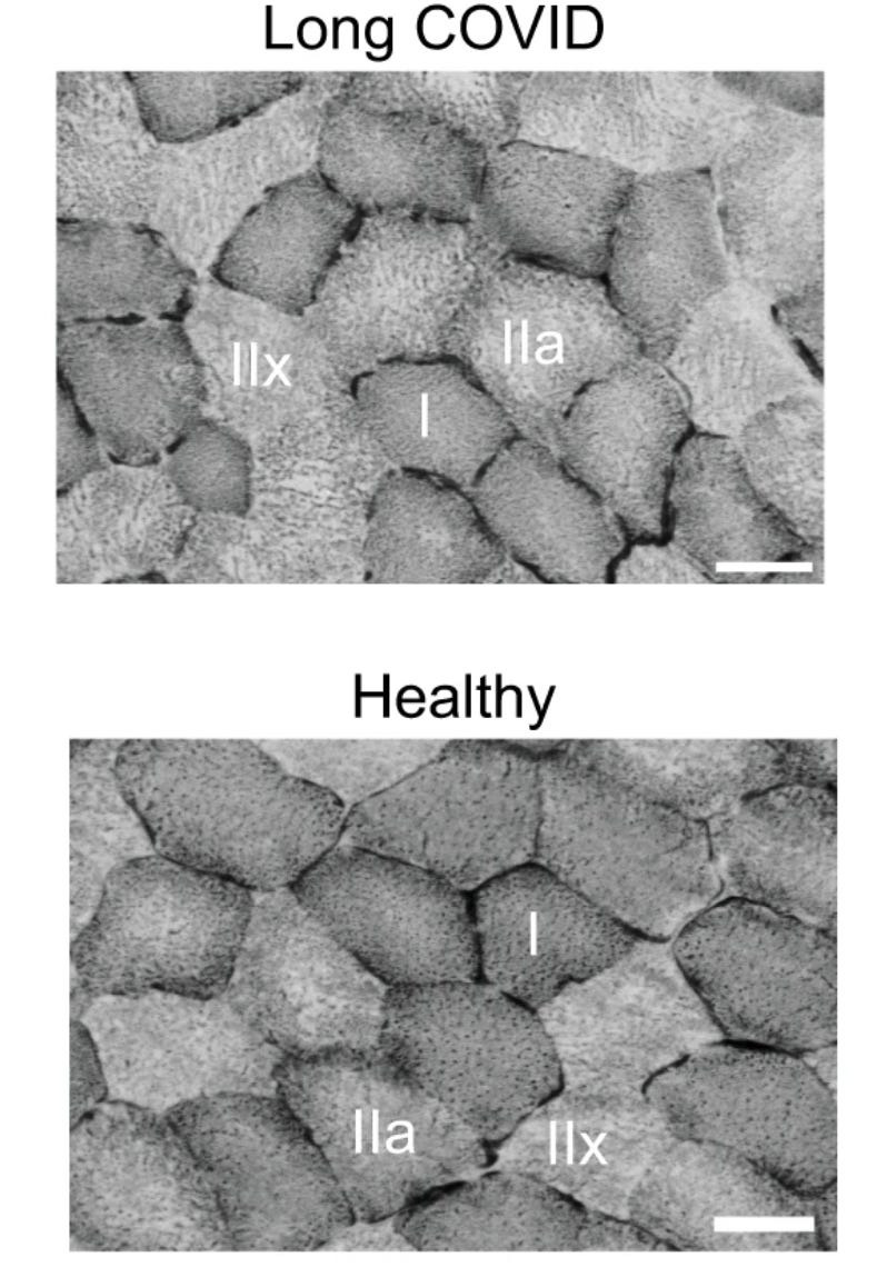
One day after exercise induced a crash, mitochondrial respiration dropped significantly in Long COVID patients while remaining stable in controls. Immune cells flooded the muscle tissue. A key energy-generating enzyme fell in patients while holding steady or rising in everyone else. Blood supply to the muscle was identical in both groups, which is the key finding: this was not oxygen-starved, deconditioned muscle. The fault was inside the cell itself, in the machinery that generates energy, and it was measurably worse after exertion. The damage was visible under a microscope. The crash is not a perception. It is a cellular event.
4. EDS: The Connective Tissue Abnormality Is Now in a Mouse
Gensemer C, Petrucci T, Beck T, et al. KLK15 alters connective tissues in hypermobile Ehlers-Danlos syndrome. iScience. 2025;28(9):113343.
Hypermobile EDS has no confirmed genetic cause, no diagnostic blood test, and until recently no animal model. Diagnosis rests entirely on clinical criteria. Patients are told their condition is genetic with no way to verify it. This study changed that in a meaningful way for the first time.
Researchers conducted whole-exome sequencing of 200 patients with hEDS and found a recurrent missense variant in a gene called KLK15 appearing across multiple unrelated families. KLK15 encodes a secreted serine protease expressed in connective tissue that interacts with components of collagen crosslinking and matrix organisation. They then created a knock-in mouse model carrying the equivalent mutation. The mice developed connective tissue abnormalities in tendons and cardiac valves, replicating hEDS features. They also showed dysregulated cytokine profiles, with significant changes in inflammatory mediators.
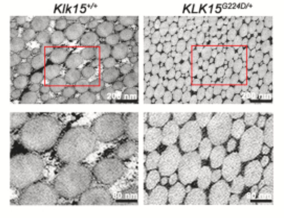
This is the first time a gene has been credibly linked to hEDS in a peer-reviewed publication with a functional animal model to support causality. A subset of hEDS patients carry a specific genetic variant that produces a specific protein abnormality that physically alters connective tissue in a way that reproduces the condition in another species. That is not a functional disorder. That is a molecular mechanism.
5. ME/CFS and Long COVID Together: The Same Biological Signature in Five Conditions
Hunter E, Alshaker H, Vugrinec D, et al. Beyond genes: EpiSwitch® and Orion platform-powered 3D genome architecture biomarkers reveal shared biology across ME/CFS, long COVID, PTSD, rheumatoid arthritis, and multiple sclerosis. Journal of Translational Medicine. 2026;24(1):1134.
I am a geneticist by day. I conduct genome-wide studies as part of my own research. So this study caught my attention in a specific way, because it approached the biology of these conditions from a layer of genomic organisation that most research does not reach: the three-dimensional architecture of the genome.
Standard genetic analysis looks at the sequence of DNA. What this study examined was how that DNA is physically folded inside cells, which regions come into contact with each other, and what those contacts mean for gene regulation. Researchers mapped these chromosome conformation signatures across ME/CFS, Long COVID, PTSD, rheumatoid arthritis and multiple sclerosis, and then asked what biological pathways were shared across all five.
The answer was striking. Despite different triggers and different clinical presentations, all five conditions showed convergence in the same regulatory networks: interferon signalling, cytokine activation including IL-1beta and TNF-alpha, mitochondrial biogenesis, cellular stress response, altered neurotransmission, glucocorticoid receptor networks governing the stress hormone axis, and VEGF signalling controlling blood vessel formation. Different conditions. Same downstream regulatory biology. The genome is folding into a similar dysregulated configuration across all of them, regardless of what caused it.
This study has limitations, including a commercial conflict of interest that should be noted given the EpiSwitch platform is a product of Oxford BioDynamics, whose employees co-authored the paper, and the fact that it requires independent replication. But the mechanistic convergence it identifies across five conditions is consistent with findings from multiple other research groups using entirely different methodologies.
Why this matters
None of these five studies proved a psychological origin for these conditions. None of them suggested that the symptoms are mediated by how patients think about their illness or how much they exercise. Each of them found something specific, measurable, and objective: abnormal blood flow in the brain, a named protein dismantling mitochondria, cellular damage visible in a biopsy, a genetic variant producing connective tissue abnormality in a mouse model, and shared dysregulation of the same biological networks across five conditions at the level of genome architecture.
These are the findings that were not supposed to exist if these conditions were functional, psychosomatic, or primarily driven by deconditioning. They exist. They are published. They are peer-reviewed. They are real.
For everyone who has been told otherwise: the science disagrees.
The views and opinions expressed in Dysautonomia Decoded are my own and do not represent those of my employer or any affiliated organisation.
References
Seeley MC, et al. Novel brain SPECT imaging unravels abnormal cerebral perfusion in POTS. Scientific Reports. 2025;15:3487. https://doi.org/10.1038/s41598-025-87748-4
Wang PY, Ma J, Kim YC, et al. WASF3 disrupts mitochondrial respiration and may mediate exercise intolerance in ME/CFS. PNAS. 2023;120(34):e2302738120. https://doi.org/10.1073/pnas.2302738120
Appelman B, Charlton BT, Goulding RP, et al. Muscle abnormalities worsen after post-exertional malaise in long COVID. Nature Communications. 2024;15:17. https://doi.org/10.1038/s41467-023-44432-3
Gensemer C, Petrucci T, Beck T, et al. KLK15 alters connective tissues in hypermobile Ehlers-Danlos syndrome. iScience. 2025;28(9):113343. https://doi.org/10.1016/j.isci.2025.113343
Hunter E, Alshaker H, Vugrinec D, et al. Beyond genes: EpiSwitch® and Orion platform-powered 3D genome architecture biomarkers reveal shared biology across ME/CFS, long COVID, PTSD, rheumatoid arthritis, and multiple sclerosis. Journal of Translational Medicine. 2026;24(1):1134. https://doi.org/10.1186/s12967-026-08874-9
THANK YOU for this groundbreaking info on hEDS actually being linked to a gene! Now if we can only get the medical community to read this!!
Thanks, a useful reference. Much appreciated.